Spring is a time when veterinarians begin to see increased cases of laminitis, a painful disease that affects the feet of horses. Laminitis results from the disruption of blood flow to the sensitive and insensitive laminae within the foot, which secure the coffin bone (P3) to the hoof wall. While the exact mechanisms by which the feet are damaged is still being determined, certain precipitating events can produce laminitis. Although laminitis occurs in the feet, the underlying cause is often a disturbance elsewhere in the horse’s body.
Recent research has led to the description of the laminae in a horse’s foot as a ‘shock organ.’ That is, an organ that fails related to some sort of systemic disease. In people, the lung is considered a “shock organ”, and they are finding that the laminae in a horse’s foot reacts very similarly. This suggests that treating other diseases more effectively — like colitis, or a mare with a retained placenta, or a horse with pleurao pneumonia — we can better protect the foot from becoming the shock organ that ultimately fails and results in a crippling disease.
Laminitis can also occur after a carbohydrate overload, where your horse eats to much rich young grass or grain. An injury or trauma that causes the horse to bear less weight on one leg may also induce laminitis, as in the case of Barbaro. The continuous increased stress placed on the weight bearing limb strains the lamina that hold the coffin bone stable in the hoof, resulting in inflammation and laminitis. It is important to provide support and cushioning to all feet and legs for a horse being treated for any injury or lameness that changes how it bears weight.
Researchers have also identified a “Pre-Laminitic Metabolic Syndrome (PLMS)” in horses similar to the metabolic syndromes in people that are considered risk factors for Type II diabetes or coronary heart disease. These horses are especially sensitive to carbohydrate overload and can ‘founder’ for no apparent reason. The researchers have used cut off points for Insulin Sensitivity, Pancreatic Beta cell Response (Insulin levels compared to glucose levels), Body Condition Score and Serum Triglyceride levels to identify these PLMS ponies which are at a higher risk of developing laminitis. Identification of PLMS in ponies (and possibly horses) would allow owners and veterinarians to manage nutrition and feeding practices appropriately to prevent laminitis prior to clinical disease.
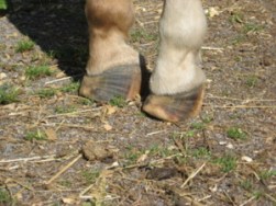
As a horse owner, it is important to recognize the signs of laminitis and seek veterinary help immediately. Signs of acute laminitis include the following:
- Lameness, especially when a horse is turning in circles; shifting lameness when standing
- Heat in the feet
- Increased digital pulse in the feet
- Pain in the toe region when pressure is applied with hoof testers
- Reluctant or hesitant gait, as if “walking on eggshells”
- A “rocking horse stance” in the front, with the front feet stretched out in front to alleviate pressure on the toes
Signs of chronic laminitis may include the following:
- Rings in hoof wall that become wider as they are followed from toe to heel
- Dished hooves, which are the result of unequal rates of hoof growth
- Thick, “cresty” neck and abnormal fat deposits (signs of metabolic disorders that often cause laminitis)
- Bruised soles or “stone bruises”
- Widened white line, commonly called “seedy toe,” with occurrence of blood pockets
and/or abscesses - Dropped soles or flat feet
If you suspect laminitis or your horse has gotten more than a normal amount of carbohydrates (grass or grain), consider it a medical emergency and notify your veterinarian immediately. Early intervention in acute laminitis carries the best prognosis. The sooner treatment begins, the better the chance for recovery.
Your veterinarian may recommend icing your horse’s feet if it is an acute case, and placing him on deep, soft bedding with no or limited grain and lower quality hay until they are able to see them. They may also recommend applying cushion/support to your horse’s feet via boots or pads made of foam insulation sheets and duct tape.
Treating laminitis requires a multi-modal approach. Your veterinarian will need to work closely with your farrier to develop a plan and identify any changes to the coffin bone-hoof structure. X-rays may be needed to look for rotation and/or sinking of the coffin bone. Close communication between your veterinarian, farrier and yourself is essential to prevent any further changes from occurring while keeping your horse comfortable during recovery.
Source Henderson Equine Clinic